Are you wondering why an echo test is done? An echocardiogram, often called an echo test, is a non-invasive ultrasound of the heart. why.edu.vn explains that this essential diagnostic tool uses sound waves to create detailed images of your heart, enabling healthcare professionals to assess its structure and function. Dive in to explore the various applications, types, and benefits of echocardiography, ensuring you’re well-informed about cardiac imaging and heart health assessment.
Table of Contents
- What is an Echocardiogram?
- How Echocardiography Works
- What an Echocardiogram Reveals
- Why Is an Echo Test Done?
- Evaluating Heart Structure and Function
- Diagnosing Heart Conditions
- Monitoring Heart Health
- Assessing the Effectiveness of Treatments
- Types of Echocardiograms
- Transthoracic Echocardiogram (TTE)
- Transesophageal Echocardiogram (TEE)
- Stress Echocardiogram
- Fetal Echocardiogram
- Intracardiac Echocardiogram (ICE)
- The Echocardiogram Procedure: What to Expect
- Preparation for the Test
- During the Procedure
- After the Procedure
- Benefits and Risks of Echocardiography
- Benefits of Echocardiography
- Risks of Echocardiography
- Understanding Your Echocardiogram Results
- Normal Findings
- Abnormal Findings
- Advancements in Echocardiography Technology
- 3D Echocardiography
- Strain Echocardiography
- Contrast Echocardiography
- The Role of Echocardiography in Specific Heart Conditions
- Heart Failure
- Valvular Heart Disease
- Congenital Heart Defects
- Cardiomyopathy
- Pericardial Disease
- Preparing for Your Echocardiogram: A Step-by-Step Guide
- Pre-Test Instructions
- What to Bring
- Questions to Ask Your Doctor
- Frequently Asked Questions (FAQs) about Echocardiograms
- Conclusion
1. What is an Echocardiogram?
An echocardiogram is a non-invasive diagnostic test that uses ultrasound technology to visualize the heart. It provides real-time images of the heart’s structure, function, and blood flow. This tool helps healthcare professionals detect abnormalities and diagnose various heart conditions. Understanding the principles behind echocardiography and what it reveals is crucial for appreciating its role in cardiac care.
How Echocardiography Works
Echocardiography employs ultrasound waves, which are high-frequency sound waves, to create images of the heart. A device called a transducer emits these sound waves, which then bounce off the different structures of the heart. The transducer receives these reflected waves, and a computer converts them into visual images displayed on a monitor. This process allows doctors to observe the heart’s movements, the size and shape of its chambers, and the function of its valves. The Doppler technique, often used in conjunction with standard echocardiography, assesses the speed and direction of blood flow, providing additional information about the heart’s performance.
What an Echocardiogram Reveals
An echocardiogram provides a wealth of information about the heart, including:
- Size and Shape of the Heart: Helps identify enlarged or thickened heart chambers, which can indicate conditions like hypertension or cardiomyopathy.
- Pumping Strength (Ejection Fraction): Measures how much blood the heart pumps out with each beat, a key indicator of heart failure.
- Function of Heart Valves: Evaluates whether the heart valves are opening and closing properly, detecting issues like stenosis (narrowing) or regurgitation (leaking).
- Blood Flow: Assesses the direction and speed of blood flow through the heart, identifying potential blockages or abnormal flow patterns.
- Presence of Blood Clots or Tumors: Detects any abnormal masses inside the heart.
- Congenital Heart Defects: Identifies structural abnormalities present from birth.
- Pericardial Effusion: Detects fluid accumulation around the heart.
 Echocardiogram transducer
Alt text: Echocardiogram being performed with a transducer, showing a healthcare professional using the device on a patient’s chest to obtain images of the heart.
2. Why Is an Echo Test Done?
An echo test, or echocardiogram, is performed for a variety of reasons, all centered around evaluating and monitoring the heart’s health. The primary reasons include assessing the heart’s structure and function, diagnosing heart conditions, monitoring the progression of heart disease, and evaluating the effectiveness of treatments.
Evaluating Heart Structure and Function
Echocardiograms are essential for visualizing the heart’s anatomy and assessing its mechanical performance. This includes:
- Chamber Size and Wall Thickness: Identifying abnormalities such as enlarged or thickened chambers, which may indicate conditions like hypertension, heart failure, or cardiomyopathy.
- Valve Function: Evaluating the opening and closing of heart valves to detect stenosis (narrowing) or regurgitation (leaking).
- Ejection Fraction (EF): Measuring the percentage of blood pumped out of the left ventricle with each contraction, a key indicator of heart failure. A normal EF is typically between 55% and 70%.
- Cardiac Output: Assessing the amount of blood the heart pumps per minute to meet the body’s needs.
- Wall Motion Abnormalities: Detecting areas of the heart muscle that are not contracting properly, which may indicate a previous heart attack or ischemia (reduced blood flow).
Diagnosing Heart Conditions
Echocardiograms play a critical role in diagnosing a wide range of heart conditions, including:
- Heart Failure: Determining the type and severity of heart failure by assessing the heart’s pumping ability and identifying structural abnormalities.
- Valvular Heart Disease: Diagnosing conditions such as aortic stenosis, mitral regurgitation, and tricuspid valve disease by visualizing valve structure and blood flow patterns.
- Congenital Heart Defects: Identifying structural abnormalities present at birth, such as atrial septal defects (ASD) or ventricular septal defects (VSD).
- Cardiomyopathy: Diagnosing various forms of cardiomyopathy, including dilated, hypertrophic, and restrictive cardiomyopathy, by assessing heart muscle thickness and function.
- Pericardial Disease: Detecting pericardial effusion (fluid around the heart) and constrictive pericarditis (thickening and stiffening of the pericardium).
- Endocarditis: Identifying vegetations (growths) on the heart valves caused by infection.
- Atrial Fibrillation: Assisting in the management of atrial fibrillation by evaluating the left atrial size and function.
Monitoring Heart Health
Regular echocardiograms are often used to monitor the progression of heart disease and assess the impact of treatments. This is particularly important for individuals with:
- Known Heart Conditions: Monitoring the stability or progression of conditions such as heart failure, valvular heart disease, and cardiomyopathy.
- Risk Factors for Heart Disease: Assessing heart health in individuals with risk factors such as hypertension, diabetes, high cholesterol, and a family history of heart disease.
- History of Heart Attack: Evaluating heart function and detecting complications after a myocardial infarction (heart attack).
- Exposure to Cardiotoxic Medications: Monitoring heart function in individuals receiving chemotherapy or other medications that can damage the heart.
Assessing the Effectiveness of Treatments
Echocardiograms are used to evaluate the effectiveness of medical and surgical interventions for heart conditions. This includes:
- Medication Management: Assessing the impact of medications on heart function in individuals with heart failure or hypertension.
- Valve Repair or Replacement: Evaluating the success of valve repair or replacement surgeries by assessing valve function and blood flow patterns.
- Cardiac Resynchronization Therapy (CRT): Assessing the effectiveness of CRT devices in improving heart function in individuals with heart failure.
- Transcatheter Aortic Valve Implantation (TAVI): Evaluating the success of TAVI procedures in replacing damaged aortic valves.
Understanding these reasons can help patients and healthcare providers make informed decisions about the need for and frequency of echocardiograms in managing heart health. Regular monitoring and timely intervention can significantly improve outcomes and quality of life for individuals with heart conditions.
3. Types of Echocardiograms
There are several types of echocardiograms, each designed to provide specific information about the heart. The choice of which type to use depends on the patient’s condition, the specific details needed, and the healthcare provider’s clinical judgment.
Transthoracic Echocardiogram (TTE)
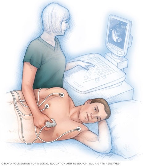
The transthoracic echocardiogram (TTE) is the most common type of echocardiogram. It is non-invasive and involves placing a transducer on the chest to obtain images of the heart.
- Procedure: The patient lies on their back or left side, and a gel is applied to the chest to improve contact between the transducer and the skin. The transducer is then moved across the chest to obtain different views of the heart.
- Uses: TTE is used to evaluate the overall structure and function of the heart, including chamber size, valve function, and ejection fraction. It is often the first test performed when evaluating heart conditions.
- Advantages: Non-invasive, readily available, and provides a comprehensive overview of heart structure and function.
- Disadvantages: Image quality can be limited by factors such as obesity, lung disease, and chest wall deformities.
Transesophageal Echocardiogram (TEE)
A transesophageal echocardiogram (TEE) provides a more detailed view of the heart than a TTE. It involves inserting a small transducer attached to the end of a flexible tube down the esophagus.
- Procedure: The patient is given a sedative to relax them, and the throat is numbed with a local anesthetic. The transducer is then guided down the esophagus, which lies directly behind the heart.
- Uses: TEE is used to evaluate conditions such as aortic valve disease, atrial fibrillation, and endocarditis. It provides high-resolution images of the heart structures, particularly the atria and valves.
- Advantages: Provides clearer images of the heart than TTE, particularly for structures that are difficult to visualize with TTE.
- Disadvantages: More invasive than TTE, requires sedation, and carries a small risk of complications such as esophageal perforation.
Stress Echocardiogram
A stress echocardiogram is used to evaluate heart function during physical activity or stress. It involves performing an echocardiogram before and immediately after exercise or pharmacological stress.
- Procedure: The patient exercises on a treadmill or stationary bike while their heart rate and blood pressure are monitored. Alternatively, a medication such as dobutamine is used to simulate the effects of exercise on the heart. Echocardiogram images are obtained before and after the stress test.
- Uses: Stress echocardiography is used to detect coronary artery disease and evaluate the heart’s response to stress. It can identify areas of the heart muscle that are not receiving enough blood flow during exercise.
- Advantages: Provides information about heart function under stress, which can help diagnose coronary artery disease.
- Disadvantages: Requires the patient to be able to exercise or tolerate pharmacological stress.
Fetal Echocardiogram
A fetal echocardiogram is a specialized ultrasound used to evaluate the heart of an unborn baby. It is typically performed during the second trimester of pregnancy.
- Procedure: The procedure is similar to a standard obstetrical ultrasound. A transducer is placed on the mother’s abdomen to obtain images of the fetal heart.
- Uses: Fetal echocardiography is used to detect congenital heart defects before birth. It can help healthcare providers plan for the baby’s care after delivery.
- Advantages: Non-invasive for the mother and provides valuable information about the fetal heart.
- Disadvantages: Image quality can be limited by factors such as maternal obesity and fetal position.
Intracardiac Echocardiogram (ICE)
An intracardiac echocardiogram (ICE) involves inserting a small transducer into the heart through a catheter. This technique is often used during electrophysiology studies or other invasive cardiac procedures.
- Procedure: A catheter with a transducer at the tip is inserted into a blood vessel (usually in the groin) and guided into the heart. The transducer then provides real-time images of the heart structures.
- Uses: ICE is used to guide and monitor complex cardiac procedures, such as atrial septal defect closures and ablation of arrhythmias.
- Advantages: Provides high-resolution images of the heart from within the heart chambers.
- Disadvantages: Invasive and carries a risk of complications such as bleeding, infection, and perforation of the heart.
Understanding the different types of echocardiograms and their specific uses can help patients and healthcare providers choose the most appropriate test for their needs. Each type of echocardiogram provides unique information that can aid in the diagnosis and management of heart conditions.
4. The Echocardiogram Procedure: What to Expect
Knowing what to expect during an echocardiogram procedure can help alleviate anxiety and ensure a smoother experience. This section outlines the typical steps involved in preparing for the test, what happens during the procedure, and what to expect afterward.
Preparation for the Test
The preparation for an echocardiogram varies depending on the type of test being performed.
- Transthoracic Echocardiogram (TTE):
- No special preparation is usually required.
- You can eat, drink, and take medications as usual unless otherwise instructed by your doctor.
- Wear comfortable clothing that allows easy access to your chest.
- Transesophageal Echocardiogram (TEE):
- You will be asked not to eat or drink for at least six hours before the test.
- Inform your doctor about any medications you are taking, especially blood thinners or diabetes medications.
- Arrange for someone to drive you home, as you will likely be given sedatives that can impair your ability to drive.
- Stress Echocardiogram:
- Avoid eating a heavy meal or drinking caffeinated beverages for a few hours before the test.
- Wear comfortable clothing and shoes suitable for exercise.
- Inform your doctor about any medications you are taking, especially beta-blockers or nitrates, as they may affect the test results.
- Fetal Echocardiogram:
- No special preparation is required.
- You may be asked to drink water before the test to improve the image quality.
During the Procedure
The echocardiogram procedure typically involves the following steps:
- Transthoracic Echocardiogram (TTE):
- You will be asked to remove your clothing from the waist up and put on a hospital gown.
- You will lie on an examination table on your back or left side.
- Electrodes (small, sticky patches) will be attached to your chest to monitor your heart’s electrical activity (ECG).
- A gel will be applied to your chest to help transmit the ultrasound waves.
- The technician will move the transducer (a small, handheld device) across your chest to obtain images of your heart.
- You may be asked to hold your breath or change positions to improve the image quality.
- The procedure usually takes about 20-60 minutes.
- Transesophageal Echocardiogram (TEE):
- You will be asked not to eat or drink for at least six hours before the test.
- An IV line will be inserted into your arm to administer sedatives.
- Your throat will be numbed with a local anesthetic spray or gel.
- A mouth guard will be placed in your mouth to protect your teeth.
- The technician will gently guide the transducer down your esophagus.
- Images of your heart will be obtained from inside your esophagus.
- Your heart rate, blood pressure, and oxygen levels will be monitored throughout the procedure.
- The procedure usually takes about 30-90 minutes.
- Stress Echocardiogram:
- You will have a resting echocardiogram performed to obtain baseline images of your heart.
- Electrodes will be attached to your chest to monitor your heart’s electrical activity (ECG).
- You will begin exercising on a treadmill or stationary bike, gradually increasing the intensity.
- Your heart rate, blood pressure, and oxygen levels will be monitored throughout the exercise.
- Echocardiogram images will be obtained immediately after you stop exercising.
- If you are unable to exercise, you may be given a medication (such as dobutamine) to simulate the effects of exercise on your heart.
- The procedure usually takes about 60-90 minutes.
- Fetal Echocardiogram:
- You will lie on an examination table.
- A gel will be applied to your abdomen.
- The technician will move the transducer across your abdomen to obtain images of the fetal heart.
- The procedure usually takes about 30-60 minutes.
After the Procedure
After the echocardiogram, you can typically resume your normal activities unless you have had a TEE.
- Transthoracic Echocardiogram (TTE):
- You can usually return to your normal activities immediately.
- There are no restrictions on eating, drinking, or taking medications.
- Transesophageal Echocardiogram (TEE):
- You will be monitored for a few hours until the sedative wears off.
- You will not be able to eat or drink until your gag reflex returns.
- Your throat may be sore for a few hours.
- Avoid driving or operating heavy machinery for at least 24 hours after the procedure.
- Stress Echocardiogram:
- You will be monitored for a short period after the test to ensure your heart rate and blood pressure return to normal.
- You can usually return to your normal activities immediately.
- Fetal Echocardiogram:
- You can usually return to your normal activities immediately.
Understanding these steps can help you feel more prepared and comfortable during your echocardiogram procedure. Always follow your doctor’s specific instructions for the best possible outcome.
5. Benefits and Risks of Echocardiography
Echocardiography is a valuable diagnostic tool with numerous benefits, but like any medical procedure, it also carries some risks. Understanding these benefits and risks can help patients make informed decisions about their care.
Benefits of Echocardiography
- Non-Invasive: Most types of echocardiograms, such as TTE and fetal echocardiography, are non-invasive, meaning they do not require any incisions or insertion of instruments into the body.
- Painless: Echocardiography is generally painless, although some patients may experience mild discomfort from the pressure of the transducer on the chest.
- Real-Time Imaging: Echocardiography provides real-time images of the heart, allowing healthcare providers to assess heart function and structure as it is happening.
- Detailed Information: Echocardiography provides detailed information about the heart’s size, shape, structure, and function, as well as blood flow patterns.
- Versatile: Echocardiography can be used to diagnose a wide range of heart conditions, including heart failure, valvular heart disease, congenital heart defects, and cardiomyopathy.
- Monitoring: Echocardiography can be used to monitor the progression of heart disease and assess the effectiveness of treatments.
- No Radiation Exposure: Unlike X-rays and CT scans, echocardiography does not involve exposure to ionizing radiation, making it safe for pregnant women and children.
- Quick and Convenient: Echocardiography procedures are generally quick and convenient, with most tests taking less than an hour to complete.
- Cost-Effective: Echocardiography is a relatively cost-effective diagnostic tool compared to other imaging modalities such as MRI and PET scans.
Risks of Echocardiography
While echocardiography is generally safe, there are some potential risks associated with certain types of the procedure.
- Transthoracic Echocardiogram (TTE):
- Mild discomfort from the pressure of the transducer on the chest.
- Rare allergic reaction to the ultrasound gel.
- Transesophageal Echocardiogram (TEE):
- Sore throat.
- Difficulty swallowing.
- Rare complications such as esophageal perforation, bleeding, or irregular heartbeats.
- Adverse reaction to sedatives.
- Stress Echocardiogram:
- Irregular heartbeats.
- Chest pain.
- Shortness of breath.
- Rare complications such as heart attack or stroke.
- Fetal Echocardiogram:
- No known risks to the mother or fetus.
- False Positives and Negatives:
- Echocardiography results may not always be accurate, leading to false positive or false negative results.
- False positive results may lead to unnecessary further testing and treatment.
- False negative results may delay diagnosis and treatment.
- Image Quality Limitations:
- Image quality may be limited by factors such as obesity, lung disease, and chest wall deformities.
Understanding these benefits and risks can help patients and healthcare providers make informed decisions about whether echocardiography is the right diagnostic tool for their needs.
6. Understanding Your Echocardiogram Results
Receiving your echocardiogram results can be a stressful time. Understanding what the results mean can help you feel more informed and prepared to discuss your heart health with your doctor. This section outlines how to interpret normal and abnormal findings on an echocardiogram.
Normal Findings
A normal echocardiogram indicates that the heart’s structure and function are within normal limits. Specific findings include:
- Normal Heart Size: The heart chambers (atria and ventricles) are of normal size. Enlarged chambers can indicate conditions like heart failure or valve disease.
- Normal Wall Thickness: The heart muscle walls are of normal thickness. Thickened walls can indicate conditions like hypertrophic cardiomyopathy or hypertension.
- Normal Ejection Fraction (EF): The ejection fraction, which measures how much blood the left ventricle pumps out with each contraction, is within the normal range (typically 55% to 70%).
- Normal Valve Function: The heart valves (mitral, aortic, tricuspid, and pulmonary) open and close properly, without any evidence of stenosis (narrowing) or regurgitation (leaking).
- Normal Blood Flow: Blood flows through the heart and blood vessels in a normal pattern, without any evidence of blockages or abnormal flow.
- No Structural Abnormalities: There are no structural abnormalities, such as congenital heart defects or tumors.
- Normal Pericardium: The pericardium (the sac surrounding the heart) is normal, without any evidence of fluid accumulation (pericardial effusion) or thickening.
Abnormal Findings
Abnormal findings on an echocardiogram may indicate a variety of heart conditions. Common abnormalities include:
- Enlarged Heart Chambers: Enlarged atria or ventricles can indicate heart failure, valve disease, or hypertension.
- Thickened Heart Walls: Thickened heart muscle walls can indicate hypertrophic cardiomyopathy or hypertension.
- Reduced Ejection Fraction (EF): A reduced ejection fraction (less than 55%) indicates that the heart is not pumping enough blood to meet the body’s needs, which can be a sign of heart failure.
- Valve Stenosis: Narrowing of a heart valve can restrict blood flow and cause symptoms such as shortness of breath and chest pain.
- Valve Regurgitation: Leaking of a heart valve can cause blood to flow backward, leading to heart failure and other complications.
- Wall Motion Abnormalities: Areas of the heart muscle that are not contracting properly can indicate a previous heart attack or ischemia (reduced blood flow).
- Congenital Heart Defects: Structural abnormalities present at birth can affect heart function and require medical or surgical intervention.
- Pericardial Effusion: Fluid accumulation around the heart can compress the heart and impair its function.
- Cardiac Tumors: Abnormal masses inside the heart can interfere with heart function and may require surgical removal.
- Aortic Aneurysm or Dissection: Enlargement or tearing of the aorta can be life-threatening and require immediate medical attention.
If your echocardiogram results are abnormal, your doctor will discuss the findings with you and recommend further testing or treatment as needed. It is important to ask questions and understand the implications of your results for your heart health.
7. Advancements in Echocardiography Technology
Echocardiography technology has advanced significantly over the years, leading to improved image quality, more detailed information, and enhanced diagnostic capabilities. These advancements include 3D echocardiography, strain echocardiography, and contrast echocardiography.
3D Echocardiography
3D echocardiography provides a three-dimensional view of the heart, allowing healthcare providers to visualize heart structures in greater detail than with traditional 2D echocardiography.
- How It Works: 3D echocardiography uses a special transducer to acquire multiple 2D images from different angles. These images are then combined using computer software to create a 3D reconstruction of the heart.
- Uses: 3D echocardiography is particularly useful for evaluating valve disease, congenital heart defects, and left ventricular function.
- Advantages: Provides a more accurate assessment of heart structures and function, improves diagnostic accuracy, and facilitates surgical planning.
- Disadvantages: Requires specialized equipment and expertise, and image quality can be affected by factors such as image noise and artifacts.
Strain Echocardiography
Strain echocardiography is a technique that measures the deformation, or “strain,” of the heart muscle during contraction. This can help detect subtle abnormalities in heart function that may not be apparent with traditional echocardiography.
- How It Works: Strain echocardiography uses sophisticated software to track the movement of the heart muscle during the cardiac cycle. This allows healthcare providers to quantify the amount of strain, or deformation, that occurs in different regions of the heart.
- Uses: Strain echocardiography is used to evaluate conditions such as heart failure, cardiomyopathy, and ischemia.
- Advantages: Detects subtle abnormalities in heart function, improves diagnostic accuracy, and provides prognostic information.
- Disadvantages: Requires specialized software and expertise, and results can be affected by factors such as image quality and heart rate.
Contrast Echocardiography
Contrast echocardiography involves injecting a contrast agent (a solution of tiny microbubbles) into the bloodstream to enhance the image quality of the echocardiogram.
- How It Works: The contrast agent travels through the bloodstream and enters the heart chambers, reflecting ultrasound waves and improving the visualization of heart structures.
- Uses: Contrast echocardiography is used to improve the visualization of the heart chambers, detect intracardiac masses, and assess myocardial perfusion (blood flow to the heart muscle).
- Advantages: Enhances image quality, improves diagnostic accuracy, and provides additional information about heart function.
- Disadvantages: Requires the injection of a contrast agent, which can cause allergic reactions in some patients.
These advancements in echocardiography technology have significantly improved the ability to diagnose and manage heart conditions. As technology continues to evolve, echocardiography is likely to play an even greater role in cardiac care.
8. The Role of Echocardiography in Specific Heart Conditions
Echocardiography is an essential diagnostic tool for a wide range of heart conditions. Its ability to provide detailed images of the heart’s structure and function makes it invaluable for diagnosing, monitoring, and managing various cardiac disorders.
Heart Failure
Echocardiography plays a crucial role in the diagnosis and management of heart failure. It can:
- Assess Left Ventricular Function: Measure the ejection fraction (EF) to determine the heart’s pumping ability. A reduced EF is a hallmark of heart failure.
- Evaluate Chamber Size and Wall Thickness: Identify enlarged or thickened heart chambers, which can indicate the underlying cause of heart failure.
- Detect Valve Abnormalities: Identify valve stenosis or regurgitation, which can contribute to heart failure.
- Assess Diastolic Function: Evaluate how well the heart relaxes and fills with blood, which is important in diagnosing diastolic heart failure.
Valvular Heart Disease
Echocardiography is essential for diagnosing and monitoring valvular heart disease, which involves abnormalities of the heart valves. It can:
- Visualize Valve Structure: Identify valve stenosis (narrowing) or regurgitation (leaking).
- Assess Valve Severity: Quantify the severity of valve stenosis or regurgitation using Doppler measurements.
- Evaluate the Impact on Heart Function: Assess how valve abnormalities are affecting the size and function of the heart chambers.
- Guide Treatment Decisions: Help determine whether medical management, valve repair, or valve replacement is necessary.
Congenital Heart Defects
Echocardiography is a primary diagnostic tool for detecting congenital heart defects, which are structural abnormalities present at birth. It can:
- Visualize Heart Structures: Identify abnormalities in the heart chambers, valves, and blood vessels.
- Assess Blood Flow Patterns: Evaluate blood flow through the heart to detect abnormal shunts or obstructions.
- Guide Interventional Procedures: Provide real-time imaging during catheter-based interventions to repair congenital heart defects.
- Monitor Post-Operative Outcomes: Assess the success of surgical repairs and identify any complications.
Cardiomyopathy
Echocardiography is used to diagnose and classify various types of cardiomyopathy, which are diseases of the heart muscle. It can:
- Assess Heart Muscle Thickness: Identify thickened or thinned heart muscle, which is characteristic of hypertrophic or dilated cardiomyopathy.
- Evaluate Heart Chamber Size and Shape: Determine whether the heart chambers are enlarged or distorted.
- Assess Left Ventricular Function: Measure the ejection fraction (EF) to assess the heart’s pumping ability.
- Detect Wall Motion Abnormalities: Identify areas of the heart muscle that are not contracting properly.
Pericardial Disease
Echocardiography is used to diagnose and monitor pericardial disease, which involves abnormalities of the pericardium (the sac surrounding the heart). It can:
- Detect Pericardial Effusion: Identify fluid accumulation around the heart.
- Assess Pericardial Thickening: Detect thickening and inflammation of the pericardium, which can indicate constrictive pericarditis.
- Evaluate the Impact on Heart Function: Assess how pericardial abnormalities are affecting the heart’s ability to fill and pump blood.
In each of these heart conditions, echocardiography provides valuable information that guides diagnosis, treatment, and management. Its non-invasive nature and ability to provide real-time images of the heart make it an indispensable tool in cardiac care.
9. Preparing for Your Echocardiogram: A Step-by-Step Guide
Preparing for an echocardiogram involves understanding the specific instructions for the type of test you are undergoing. Proper preparation can ensure accurate results and a smooth experience.
Pre-Test Instructions
- Consult Your Doctor: Discuss any medications you are taking, including prescription drugs, over-the-counter medications, and supplements. Your doctor will advise you on whether to continue or temporarily stop taking any medications before the test.
- Fasting Instructions: For a transesophageal echocardiogram (TEE), you will typically need to fast for at least six hours before the test. Follow your doctor’s specific instructions regarding food and drink.
- Hydration: For a transthoracic echocardiogram (TTE) or fetal echocardiogram, you can usually eat, drink, and take medications as usual. However, your doctor may advise you to drink water before the test to improve image quality.
- Avoid Caffeine and Alcohol: For a stress echocardiogram, avoid caffeine and alcohol for at least 24 hours before the test, as they can affect your heart rate and blood pressure.
- Arrange Transportation: If you are having a TEE, arrange for someone to drive you home, as you will likely receive sedation that can impair your ability to drive.
- Wear Comfortable Clothing: Wear comfortable clothing that allows easy access to your chest. You may be asked to change into a hospital gown for the test.
What to Bring
- Insurance Information: Bring your insurance card and any required paperwork.
- Identification: Bring a valid form of identification, such as a driver’s license or passport.
- Medication List: Bring a list of all medications you are taking, including dosages and frequency.
- Doctor’s Orders: Bring any written orders or instructions from your doctor.
- Comfort Items: Consider bringing a book, magazine, or music to help you relax during the waiting period.
Questions to Ask Your Doctor
- Why do I need an echocardiogram? Understanding the reason for the test can help you feel more informed and involved in your care.
- What type of echocardiogram will I be having? Knowing the type of test will help you understand the specific preparation instructions.
- How should I prepare for the test? Ask about any specific instructions regarding food, drink, medications, and activities.
- What are the risks and benefits of the test? Understanding the potential risks and benefits can help you make an informed decision about whether to undergo the test.
- What will happen during the test? Asking about the procedure can help alleviate anxiety and ensure a smoother experience.
- How long will the test take? Knowing the duration of the test can help you plan your day accordingly.
- When will I receive the results? Ask when you can expect to receive the results and how they will be communicated to you.
- What happens after the test? Ask about any post-test instructions or follow-up appointments.
By following these steps and asking the right questions, you can prepare for your echocardiogram and ensure a smooth and informative experience.
10. Frequently Asked Questions (FAQs) about Echocardiograms
Here are some frequently asked questions about echocardiograms, along with detailed answers to help you better understand the procedure.
Q1: What is an echocardiogram?
A: An echocardiogram is a non-invasive diagnostic test that uses ultrasound waves to create images of the heart. It provides real-time information about the heart’s structure, function, and blood flow.
Q2: Why do I need an echocardiogram?
A: An echocardiogram is performed to evaluate the heart’s structure and function, diagnose heart conditions, monitor the progression of heart disease, and assess the effectiveness of treatments.
Q3: What are the different types of echocardiograms?
A: The main types of echocardiograms include:
- Transthoracic Echocardiogram (TTE)
- Transesophageal Echocardiogram (TEE)
- Stress Echocardiogram
- Fetal Echocardiogram
- Intracardiac Echocardiogram (ICE)
Q4: How should I prepare for an echocardiogram?
A: Preparation depends on the type of echocardiogram. Generally:
- For a TTE, no special preparation is needed.
- For a TEE, you will need to fast for at least six hours before the test.
- For a stress echocardiogram, avoid caffeine and alcohol for at least 24 hours before the test.
Q5: Is an echocardiogram painful?
A: An echocardiogram is generally painless. You may experience mild discomfort from the pressure of the transducer on the chest during a TTE. A TEE may cause a sore throat afterward.
Q6: How long does an echocardiogram take?
A: The duration of an echocardiogram varies depending on the type of test:
- TTE: 20-60 minutes
- TEE: 30-90 minutes
- Stress Echocardiogram: 60-90 minutes
- Fetal Echocardiogram: 30-60 minutes
Q7: What are the risks of an echocardiogram?
A: Echocardiography is generally safe. Risks vary depending on the type of test:
- TTE: Minimal risks, such as mild discomfort.
- TEE: Sore throat, difficulty swallowing, rare complications such as esophageal perforation.
- Stress Echocardiogram: Irregular heartbeats, chest pain, rare complications such as heart attack.
Q8: How will I receive my echocardiogram results?
A: Your doctor will review the echocardiogram results and discuss them with you during a follow-
