Gel electrophoresis is a cornerstone technique in molecular biology and genetics, used to separate DNA fragments based on their size. You might have seen images of DNA bands neatly lined up in gels, but have you ever stopped to consider why DNA migrates through this gel and, more specifically, why it moves towards the positive electrode? This article will explain the fundamental principle behind this movement and why it’s crucial for DNA analysis.
The Intrinsic Negative Charge of DNA
The key to understanding DNA’s movement in gel electrophoresis lies in its inherent electrical charge. DNA, or deoxyribonucleic acid, is composed of repeating units called nucleotides. Each nucleotide consists of a deoxyribose sugar, a phosphate group, and a nitrogenous base. It’s the phosphate group that carries a negative charge.
Each phosphate group in the DNA backbone has a negatively charged oxygen atom. Since DNA is made up of many nucleotides linked together, it carries an overall negative charge proportional to its length. This negative charge is a fundamental property of DNA and is consistent regardless of the sequence of bases.
Setting Up the Electric Field in Gel Electrophoresis
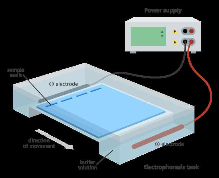
Gel electrophoresis utilizes an electric field to drive the movement of molecules. The setup involves a gel, typically made of agarose or polyacrylamide, submerged in a buffer solution within an electrophoresis tank. An electric current is then applied across the gel, creating a defined positive and negative electrode.
Think of it like setting up a battery circuit. One end of the electrophoresis tank is connected to the positive terminal, and the other end to the negative terminal. This creates an electrical field that runs through the gel matrix.
Opposites Attract: DNA’s Migration to the Positive Pole
Now, let’s bring together the negative charge of DNA and the electric field. A basic principle of physics is that opposite charges attract. Since DNA is negatively charged, it will be attracted to the positive electrode in the electrophoresis tank.
When the electric current is switched on, the negatively charged DNA molecules experience a force that pulls them towards the positive electrode (also called the anode). This is the fundamental reason why DNA moves to the positive electrode during gel electrophoresis. It’s a direct consequence of the electrostatic attraction between opposite charges.
Image alt text: Gel electrophoresis apparatus setup, illustrating DNA samples in wells of a gel submerged in buffer within a tank, connected to a power supply with positive and negative electrodes.
Size-Based Separation in the Gel Matrix
While the electric field drives DNA towards the positive electrode, the gel matrix itself plays a crucial role in separating DNA fragments by size. The gel acts like a sieve, a porous material with tiny channels running through it.
When DNA fragments move through the gel, shorter fragments encounter less resistance and can navigate through the pores more easily. Consequently, they travel faster and further towards the positive electrode. Conversely, longer DNA fragments experience more resistance, move more slowly, and travel a shorter distance in the same amount of time.
This difference in migration speed based on size is what allows gel electrophoresis to separate DNA fragments. After running the gel, you’ll find smaller DNA fragments closer to the positive electrode and larger fragments further away, creating distinct bands.
Visualizing Separated DNA Bands
After electrophoresis, the separated DNA fragments within the gel are not visible to the naked eye. To see them, a DNA staining dye is used. This dye binds to DNA and fluoresces under ultraviolet (UV) light, making the DNA bands visible as bright patterns.
By comparing the positions of these bands to a DNA marker (also known as a DNA ladder) which contains fragments of known sizes, scientists can determine the approximate sizes of the DNA fragments in their samples. This is essential for various applications, from DNA fingerprinting to gene cloning and diagnostic testing.
Image alt text: Image depicting DNA bands separated on an agarose gel after electrophoresis, showing distinct bands and a DNA marker for size comparison.
The Importance of Positive Electrode Migration
The fact that DNA migrates towards the positive electrode is not just a curious phenomenon; it’s the very foundation of gel electrophoresis as a separation technique. This directed movement, driven by the fundamental charge property of DNA, allows us to:
- Separate DNA fragments by size: Essential for analyzing DNA structure, gene mapping, and genetic engineering.
- Visualize DNA: Staining allows us to see the separated fragments and analyze their patterns.
- Quantify DNA: The intensity of bands can be used to estimate the amount of DNA present.
In conclusion, DNA moves to the positive electrode in gel electrophoresis because DNA is inherently negatively charged due to its phosphate backbone. This negative charge interacts with the electric field, causing DNA to migrate towards the positive pole. Coupled with the sieving effect of the gel matrix, this principle enables the powerful separation of DNA fragments by size, making gel electrophoresis an indispensable tool in biological research and diagnostics.