Why Do Cells Need To Divide? Cell division is fundamental for life, enabling growth, repair, and reproduction. At WHY.EDU.VN, we provide clear, expert-backed explanations of complex biological processes like cell division, offering solutions to your learning needs. Explore cellular replication, mitosis, and meiosis for a comprehensive understanding.
1. Understanding the Necessity of Cell Division
Cell division is a critical process that allows organisms to grow, repair tissues, and reproduce. Without it, life as we know it would not be possible. This section delves into the fundamental reasons why cells must divide, exploring the limitations of cell size and the essential functions that division facilitates.
1.1. Growth and Development
Multicellular organisms begin as a single cell, the zygote, formed through fertilization. The development from a single cell to a complex organism with trillions of cells relies entirely on cell division.
- Cell Proliferation: The increase in the number of cells through division is essential for growth. As an organism grows, more cells are needed to form tissues and organs.
- Differentiation: Cell division is often coupled with differentiation, where cells specialize to perform specific functions. For example, stem cells divide and differentiate into various cell types like muscle cells, nerve cells, or blood cells. According to research published in “Nature,” understanding the mechanisms of cell differentiation can provide insights into developmental biology and regenerative medicine.
1.2. Repair and Regeneration
Cell division plays a vital role in repairing damaged tissues and regenerating lost body parts in some organisms.
- Wound Healing: When tissues are injured, cells divide to replace the damaged or dead cells. For instance, skin cells divide rapidly to close a wound. Growth factors and cytokines stimulate this process, as discussed in “The Journal of Cell Biology.”
- Organ Regeneration: Certain animals, like salamanders, can regenerate entire limbs. This remarkable ability depends on cell division, where cells at the site of amputation divide and differentiate to rebuild the missing limb. Research in “Developmental Biology” explores the molecular mechanisms underlying regeneration.
1.3. Reproduction
Cell division is the basis for both asexual and sexual reproduction.
- Asexual Reproduction: In unicellular organisms like bacteria and yeast, cell division is the primary mode of reproduction. A single cell divides into two identical daughter cells through binary fission or mitosis.
- Sexual Reproduction: In multicellular organisms, sexual reproduction involves the fusion of gametes (sperm and egg cells) produced through meiosis, a specialized type of cell division that halves the chromosome number. According to “Genetics,” meiosis ensures genetic diversity by shuffling genes during gamete formation.
1.4. Maintaining Optimal Cell Size
Cells need to stay small to maintain an efficient surface area-to-volume ratio. This ratio affects the cell’s ability to transport nutrients and waste products across its membrane.
- Surface Area to Volume Ratio: As a cell grows, its volume increases more rapidly than its surface area. The plasma membrane, representing the surface area, is responsible for nutrient intake and waste removal. If the cell becomes too large, the surface area becomes insufficient to support the volume, leading to starvation and waste buildup.
- Diffusion Efficiency: Nutrients and waste products move through the cell via diffusion. Diffusion is more efficient over short distances. Large cells increase the time it takes for substances to diffuse, hindering cellular processes.
- Nuclear Control: The nucleus controls cellular activities. As cell volume increases, it becomes challenging for the nucleus to manage cellular processes effectively.
- Supporting Research: According to research in “Cell Biology,” the surface area-to-volume ratio is a fundamental constraint on cell size, influencing cell division and specialization.
2. The Orchestration of Cell Division: The Cell Cycle
The cell cycle is a highly regulated sequence of events that includes cell growth, DNA replication, and cell division. This section provides an in-depth overview of the cell cycle, highlighting the key phases and regulatory mechanisms that ensure accurate and timely division.
2.1. Overview of the Cell Cycle
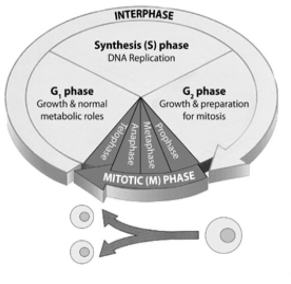
The cell cycle consists of two major phases: interphase and the mitotic (M) phase. Interphase is the period between cell divisions, during which the cell grows and prepares for division. The M phase includes mitosis (nuclear division) and cytokinesis (cytoplasmic division).
- Interphase: This phase is subdivided into G1, S, and G2 phases.
- G1 Phase (First Gap): The cell grows in size, synthesizes proteins and organelles, and performs its normal functions. The G1 phase is also a critical decision point where the cell determines whether to divide, delay division, or enter a resting state (G0 phase).
- S Phase (Synthesis): DNA replication occurs, resulting in two identical copies of each chromosome (sister chromatids). The centrosome is also duplicated during this phase.
- G2 Phase (Second Gap): The cell continues to grow and synthesizes proteins necessary for cell division. It also checks the replicated DNA for errors and repairs any damage.
- M Phase (Mitotic Phase): This phase includes mitosis and cytokinesis.
- Mitosis: The process of nuclear division, resulting in two identical nuclei. Mitosis is divided into five stages: prophase, prometaphase, metaphase, anaphase, and telophase.
- Cytokinesis: The division of the cytoplasm, resulting in two separate daughter cells.
2.2. Detailed Look at Interphase
Interphase is a crucial period of preparation for cell division. Each subphase has specific functions that ensure the cell is ready for accurate division.
- G1 Phase Activities: During G1, the cell actively engages in protein synthesis, organelle production, and metabolic activities. The cell’s fate is determined during this phase, influenced by growth factors, nutrient availability, and cell size.
- S Phase Replication: DNA replication is a highly accurate process that ensures each daughter cell receives an identical copy of the genome. Enzymes like DNA polymerase and ligase play key roles in this process. Errors during replication can lead to mutations, which can have detrimental effects on the cell.
- G2 Phase Preparations: The G2 phase allows the cell to correct any DNA replication errors and synthesize proteins required for mitosis, such as tubulin, which forms microtubules of the mitotic spindle. The cell also accumulates energy reserves needed for cell division.
2.3. Mitosis: Dividing the Nucleus
Mitosis is a precise and coordinated process that ensures each daughter cell receives an identical set of chromosomes. The process is divided into distinct stages, each with specific events.
- Prophase: The first stage of mitosis, during which the chromatin condenses into visible chromosomes. The nuclear envelope breaks down, and the mitotic spindle begins to form from the centrosomes.
- Prometaphase: The nuclear envelope completely disappears, and the spindle microtubules attach to the kinetochores of the chromosomes. The kinetochore is a protein structure located at the centromere of each chromosome.
- Metaphase: The chromosomes align along the metaphase plate, an imaginary plane equidistant from the two spindle poles. The mitotic spindle ensures that each chromosome is correctly attached to microtubules from opposite poles.
- Anaphase: The sister chromatids separate and are pulled towards opposite poles of the cell by the shortening of the spindle microtubules. Each chromatid is now considered an individual chromosome.
- Telophase: The final stage of mitosis, during which the chromosomes arrive at the poles, and the nuclear envelope reforms around each set of chromosomes. The chromosomes begin to decondense, and the mitotic spindle disappears.
2.4. Cytokinesis: Dividing the Cytoplasm
Cytokinesis is the process of dividing the cytoplasm to form two separate daughter cells. The mechanism of cytokinesis differs in animal and plant cells.
- Animal Cell Cytokinesis: A cleavage furrow forms at the middle of the cell, which deepens and eventually pinches the cell into two. The cleavage furrow is formed by a contractile ring of actin filaments and myosin.
- Plant Cell Cytokinesis: A cell plate forms at the middle of the cell, which expands and eventually fuses with the existing cell wall, separating the two daughter cells. The cell plate is formed by vesicles containing cell wall material.
3. The Significance of Meiosis in Sexual Reproduction
Meiosis is a specialized type of cell division that reduces the chromosome number by half, producing haploid gametes (sperm and egg cells). This section explores the unique features of meiosis and its importance in sexual reproduction and genetic diversity.
3.1. Meiosis I: Separating Homologous Chromosomes
Meiosis I is the first division in meiosis, during which homologous chromosomes are separated. This division includes prophase I, metaphase I, anaphase I, and telophase I.
- Prophase I: The most complex phase of meiosis, during which homologous chromosomes pair up and form tetrads. Crossing over occurs, where genetic material is exchanged between homologous chromosomes, increasing genetic diversity.
- Metaphase I: The tetrads align along the metaphase plate.
- Anaphase I: Homologous chromosomes separate and move towards opposite poles.
- Telophase I: Chromosomes arrive at the poles, and the cell divides, resulting in two haploid cells.
3.2. Meiosis II: Separating Sister Chromatids
Meiosis II is similar to mitosis, during which sister chromatids are separated. This division includes prophase II, metaphase II, anaphase II, and telophase II.
- Prophase II: Chromosomes condense, and the nuclear envelope breaks down.
- Metaphase II: Chromosomes align along the metaphase plate.
- Anaphase II: Sister chromatids separate and move towards opposite poles.
- Telophase II: Chromosomes arrive at the poles, and the cell divides, resulting in four haploid cells.
3.3. The Role of Meiosis in Genetic Diversity
Meiosis plays a crucial role in generating genetic diversity through crossing over and independent assortment of chromosomes.
- Crossing Over: The exchange of genetic material between homologous chromosomes during prophase I results in new combinations of genes.
- Independent Assortment: The random alignment of homologous chromosomes during metaphase I results in different combinations of chromosomes in the daughter cells.
- Fertilization: The fusion of two haploid gametes during fertilization restores the diploid chromosome number and creates a genetically unique individual.
4. Regulation of Cell Division: Ensuring Accuracy and Control
Cell division is tightly regulated to ensure accurate replication and segregation of chromosomes. This section explores the various checkpoints and regulatory proteins that control the cell cycle and prevent errors.
4.1. Cell Cycle Checkpoints
Cell cycle checkpoints are control mechanisms that ensure the cell cycle progresses correctly. These checkpoints monitor the integrity of DNA, the completion of DNA replication, and the proper attachment of chromosomes to the mitotic spindle.
- G1 Checkpoint: This checkpoint determines whether the cell should proceed to the S phase. Factors such as cell size, nutrient availability, and DNA damage are assessed. If conditions are unfavorable, the cell may enter the G0 phase or undergo apoptosis (programmed cell death).
- G2 Checkpoint: This checkpoint ensures that DNA replication is complete and that any DNA damage has been repaired before the cell enters mitosis.
- M Checkpoint (Spindle Checkpoint): This checkpoint ensures that all chromosomes are correctly attached to the mitotic spindle before the sister chromatids separate during anaphase.
4.2. Regulatory Proteins: Cyclins and CDKs
Cyclins and cyclin-dependent kinases (CDKs) are key regulatory proteins that control the cell cycle.
- Cyclins: These proteins fluctuate in concentration during the cell cycle and bind to CDKs, activating them.
- CDKs: These are enzymes that phosphorylate target proteins, regulating their activity and driving the cell cycle forward.
4.3. Growth Factors and Cell Communication
Growth factors are external signals that stimulate cell division. These factors bind to receptors on the cell surface, triggering signaling pathways that promote cell cycle progression.
- Mitogens: These growth factors stimulate cell division by overcoming cell cycle checkpoints.
- Inhibitory Signals: Some signals inhibit cell division, preventing uncontrolled proliferation.
5. Consequences of Uncontrolled Cell Division: Cancer
Uncontrolled cell division is a hallmark of cancer. Mutations in genes that regulate the cell cycle can lead to uncontrolled proliferation, invasion of tissues, and metastasis. This section explores the genetic and cellular mechanisms underlying cancer development.
5.1. Genetic Mutations and Cancer
Mutations in genes that control cell division, DNA repair, and apoptosis can lead to cancer.
- Proto-oncogenes: These genes promote cell division. Mutations that increase the activity of proto-oncogenes can transform them into oncogenes, which drive uncontrolled cell proliferation.
- Tumor Suppressor Genes: These genes inhibit cell division and promote apoptosis. Mutations that inactivate tumor suppressor genes can lead to uncontrolled cell proliferation.
5.2. Hallmarks of Cancer
Cancer cells exhibit several distinctive characteristics that contribute to their uncontrolled growth and spread.
- Sustaining Proliferative Signaling: Cancer cells can produce their own growth factors or activate signaling pathways that promote cell division.
- Evading Growth Suppressors: Cancer cells can inactivate tumor suppressor genes, allowing them to bypass normal growth controls.
- Resisting Cell Death: Cancer cells can evade apoptosis, allowing them to survive and proliferate even when damaged.
- Enabling Replicative Immortality: Cancer cells can reactivate telomerase, an enzyme that maintains telomere length, allowing them to divide indefinitely.
- Inducing Angiogenesis: Cancer cells can stimulate the formation of new blood vessels, providing them with nutrients and oxygen.
- Activating Invasion and Metastasis: Cancer cells can invade surrounding tissues and spread to distant sites in the body.
5.3. Cancer Therapies
Cancer therapies aim to target the unique characteristics of cancer cells, such as their uncontrolled proliferation and resistance to cell death.
- Chemotherapy: Uses drugs that kill rapidly dividing cells.
- Radiation Therapy: Uses high-energy radiation to damage cancer cells.
- Targeted Therapy: Uses drugs that target specific molecules or pathways involved in cancer cell growth and survival.
- Immunotherapy: Uses the body’s own immune system to fight cancer.
6. Common Questions About Cell Division
To further assist your understanding, here are some frequently asked questions regarding cell division.
6.1. FAQ on Cell Division
- Why do cells need to divide instead of just growing bigger?
- Cells divide to maintain an efficient surface area-to-volume ratio for nutrient and waste transport.
- What are the main phases of the cell cycle?
- The cell cycle includes interphase (G1, S, G2 phases) and the mitotic (M) phase (mitosis and cytokinesis).
- What happens during mitosis?
- Mitosis is the process of nuclear division, resulting in two identical nuclei through stages: prophase, prometaphase, metaphase, anaphase, and telophase.
- How does cytokinesis differ in animal and plant cells?
- In animal cells, cytokinesis involves the formation of a cleavage furrow, while in plant cells, it involves the formation of a cell plate.
- What is meiosis, and why is it important?
- Meiosis is a specialized type of cell division that reduces the chromosome number by half, producing haploid gametes for sexual reproduction and promoting genetic diversity.
- What are cell cycle checkpoints, and why are they important?
- Cell cycle checkpoints are control mechanisms that ensure the cell cycle progresses correctly by monitoring DNA integrity, replication, and chromosome attachment.
- What are cyclins and CDKs, and how do they regulate the cell cycle?
- Cyclins and cyclin-dependent kinases (CDKs) are key regulatory proteins that control the cell cycle by phosphorylating target proteins and driving the cell cycle forward.
- How can uncontrolled cell division lead to cancer?
- Mutations in genes that regulate the cell cycle, DNA repair, and apoptosis can lead to uncontrolled cell proliferation, invasion of tissues, and metastasis, resulting in cancer.
- What are the hallmarks of cancer cells?
- Hallmarks of cancer cells include sustaining proliferative signaling, evading growth suppressors, resisting cell death, enabling replicative immortality, inducing angiogenesis, and activating invasion and metastasis.
- What are some common cancer therapies?
- Common cancer therapies include chemotherapy, radiation therapy, targeted therapy, and immunotherapy.
7. Conclusion: The Indispensable Role of Cell Division
Cell division is essential for life, enabling growth, repair, reproduction, and genetic diversity. Understanding the intricacies of cell division is crucial for advancing our knowledge of biology and developing new therapies for diseases like cancer.
At WHY.EDU.VN, we are committed to providing you with accurate, comprehensive, and understandable explanations of complex biological processes. Whether you’re a student, a researcher, or simply curious about the world around you, we are here to help you explore the fascinating world of cell division and beyond.
Do you have more questions about cell division or any other topic? Visit why.edu.vn at 101 Curiosity Lane, Answer Town, CA 90210, United States, or contact us via Whatsapp at +1 (213) 555-0101 to ask your questions and receive expert answers. Let us help you unlock the mysteries of science and expand your knowledge.